How to improve thoracic spine mobility?
Thoracic spine mobility is a hot topic among sports coaches, Pilates and Yoga instructors.
A lack of mobility in this region is often linked to:
Poor posture: forward head posture, rounded shoulders, rounded back
Poor mobility in the upper limbs: there will be difficulty to fully flex the shoulder when doing shoulder presses or anything involving overhead movements
Different pathologies: headache, tension headaches, muscles' tension in the neck and shoulders, thoracic outlet syndrome, shoulder impingement (sub-acromial impingement)
Physiological consequences: blood and lymph circulation, decreased respiration capacity.
The purpose of this article is to:
Briefly describe the structures and functions of the thorax
Explain the dysfunctions linked to a lack of thoracic mobility based on Singla & Veqar (2017) medical study [1]
Provide a series of exercises to help you gain optimal thoracic mobility
Thorax anatomy:
The structure is formed by the T1 to T12 vertebrae's, the rib cage, the intercostal muscles and the diaphragm. The thoracic structure serves as an attachment to the upper limbs, back, neck and abdomen muscles. The thorax is also a region where respiratory mechanisms, rib movements, and lymphatic drainage and venous return occur (1).
The anatomy of the thorax is interesting because of its contradiction. Despite the 150 joints that move each time you inhale, this structure remains relatively rigid due to its attachment to the rib cage (2) (Barral, 1991). The thorax aims to protect the lungs and the heart while allowing those organs to function with optimal mobility and motility.
1. Spine
The vertebral arrangement of the thoracic spine is fascinating. The curve is made up of 12 vertebrae. The vertebrae's spinous process (central ridge of the vertebrae) T1 to T7 lengthens to serve as a continuation of the cervical vertebrae that precede them. The shape of the last thoracic vertebrae's T11 and T12 resembles that of the first lumbar L1 (3).
Like the cervical and lumbar spines, the intervertebral discs act as shock absorbers.
A healthy thoracic spine has a kyphotic curve allowing the body to resist compressive forces (4). It is important to mention that an excess of this curve may reduce breathing capacity.
Because of the rib cage, the thoracic spine's flexion and extension are somewhat limited. The degree of rotation of the thoracic curvature is 30°–35°. It is nevertheless essential to maintain this rotation in its full range of motion. When the thoracic rotation is restricted, the lumbar curve is forced to compensate. This compensation can lead to back pain or injury as the lumbar spine only tolerates 5°–10° of rotation.
2. The ribcage
The ribcage is formed by twelve pairs of ribs and their cartilage, the intercostal muscles, and the sternum (breastbone).
The ribs:
There are three different types of ribs: the true ribs, the false ribs and the floating ribs.
Ribs 1 through 7 are the "true ribs" because they attach directly to the breastbone.
The "false ribs" (ribs 8, 9 and often 10) share joint cartilage that connects them to the sternum.
The floating ribs (ribs 11 and 12) have no attachment with the sternum (1).
The intercostal muscles:
Internal and external intercostal muscles are essential for breathing. The internals elevate the rib cage and allow inhalation. The externals intercostal lower the rib cage and contribute to exhalation (5).
The sternum:
The breastbone is a flat bone that connects to the clavicle and the rib cage. It serves as a muscle attachment for the diaphragm and rectus abdominis (the 6 packs).
3. The diaphragm
The diaphragm is a musculotendinous structure that separates the thorax from the abdomen.
It works as a pump between these two regions by alternately varying their pressure. When the diaphragm contracts, the thoracic pressure decreases and the intra-abdominal pressure increases. When the diaphragm relaxes, the intra-abdominal pressure decreases, and thoracic pressure increases.
This mechanism is important for three reasons: respiration, venous return and lymphatic drainage.
During inspiration, the diaphragm contracts and the thorax's diameter increases so that air can fill the lungs. This concept of pressure is used in weightlifting. During inspiration, the intra-abdominal pressure is high enough to support the spine and prevent it from flexing (6).
The intra-abdominal pressure promotes venous return from the inferior vena cava to the right atrium of the heart. It also stimulates the passage of lymph upward through the thoracic duct.
From an osteopathic point of view, a bone or soft tissue restriction in the thorax may affect not only its entire structure but also:
Generate lower back pain
Affect the mobility of the upper limbs
Affect the venous return or the lymphatic system
Reversely, a dysfunction of the structures mentioned above may have consequences on the thoracic structure. (Barral, 1991) (2).
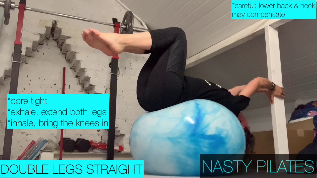
The video below suggests a series of exercises to improve the mobility of the thorax.
Impact of thoracic postural dysfunctions
What is good posture?
A good posture follows the centre of gravity line passing through the mastoid process – the shoulder – the hip – the knee, and the ankle joint. This line is used as a reference from which we can notice any deviation such as forward shoulders, forward head posture, excessive kyphosis.
1. Rounded shoulder posture:
Causes:
Using computers, tablets, and cell phones for an extended time
Seating at the desk
Carrying backpack
Repetitive overhead shoulder movement non involving a full range of motion
Rounded shoulder posture is typically associated with short pectoral minor and major muscles and overstretched rhomboids, mid and lower trapezius. Tensions may appear in the upper trapezius.
This muscles imbalance leads to an anterior rotation of the humerus and modifies the scapula's position (shoulder blades). The scapula elevates, adducts and rotate internally. It is commonly seen it winging, giving the impression of a chicken wing.
These biomechanicals changes may lead to:
Shoulder pain
Crepitus
Sub-acromial impingement (shoulder impingement)
Thoracic outlet syndrome
Arthritis in the long term.
Neck pain
Pins and needles, numbness due to the compressions of the nerves
Reduced range of motion
Reduced muscles strength and performances
2. Forward head posture
Causes:
Using computers, tablets, and cell phones for an extended period of time
Seating at the desk
Carrying backpack
Overused shoulders
In this posture, upper traps, sternocleidomastoid and levator scapula are getting shorter. This shortening of the muscles compresses the cervical vertebrae's and increase their curvature.
These biomechanicals changes may lead to:
Chronic tensions in the neck area
Headaches
In the long term, spondylosis due to the compression of the cervical spine
Shoulder pain
Temporal-mandibular disorder
Thoracic outlet syndrome
Sub-acromial (shoulder) impingement
Reduced neck mobility
Reduced breathing capacity
Hyperkyphosis
Hyperkyphosis is an increased curve of the thoracic spine which causes around back posture.
The shortening of the anterior longitudinal ligament and the upper abdominals muscles lead to a compression of the thoracic anterior vertebrae's. Posterior longitudinal ligament and extensors muscles are overstretched.
Hyperkyphosis is often seen with other postural dysfunctions such as a forward head posture and or lordosis.
Untreated hyperkyphosis can leads to:
Lower back pain
Osteoporosis
Decreased respiratory capacity
Conclusion
This article has briefly described the thorax's structures and functions and explained how they affect respiration, the lumbar spine and lymphatic and venous systems. Postural deviations arising from poor thoracic mobility are forward head posture, rounded shoulders and hyperkyphosis. Studies show correlations between those 3 deviations.
From an osteopathic point of view, a bone or soft tissue restriction in the thorax may affect not only its entire structure but also:
Generate lower back pain
Affect the mobility of the upper limbs
Affect the venous return or the lymphatic system
Reversely, a dysfunction of the structures mentioned above may have consequences on the thoracic structure. (Barral, 1991) (2).
References
1. Singla, D., Veqar, Z. (2017). Association Between Forward Head, Rounded Shoulders, and Increased Thoracic Kyphosis: A Review of the Literature. Journal of chiropractic medicine, 16(3), 220–229. https://doi.org/10.1016/j.jcm.2017.03.004
2. Moore, K., Dalley, A. & Agur, A. (2014). Clinical orientated anatomy. International Edition: Wolters Kluwer.
3. Barral, J.P. (1991). The thorax. Eastland Press Incorporated, Seattle, Washington, USA.
4. College of Osteopaths. (2021) Form & Function. The thorax.
5. Verssier, G. Biomécanique du rachis. [online]. Available at : (http://www.clubortho.fr/cariboost_files/cours_20biomecanique_20rachis_20GV.pdf). (Access March 15th 2021).
6. Biel, A. (2005). Trail guide to the body. Third Edition. Book of discovery.
7. Snell, R. (2004). Clinical anatomy. 4th edition. Lippincott Williams & Wilkins.